Tag Archive for: skin screening

Skin Cancer Myths

By Caroline Glick, PA-C, MPH


In the United States, skin cancer is the most common type of cancer. One in five Americans will develop skin cancer in their lifetime. Despite such high numbers of skin cancer diagnoses, there are many myths pertaining to skin cancer and sun protection. In this post, I will debunk some of these common myths.

Common Skin Cancer Myths

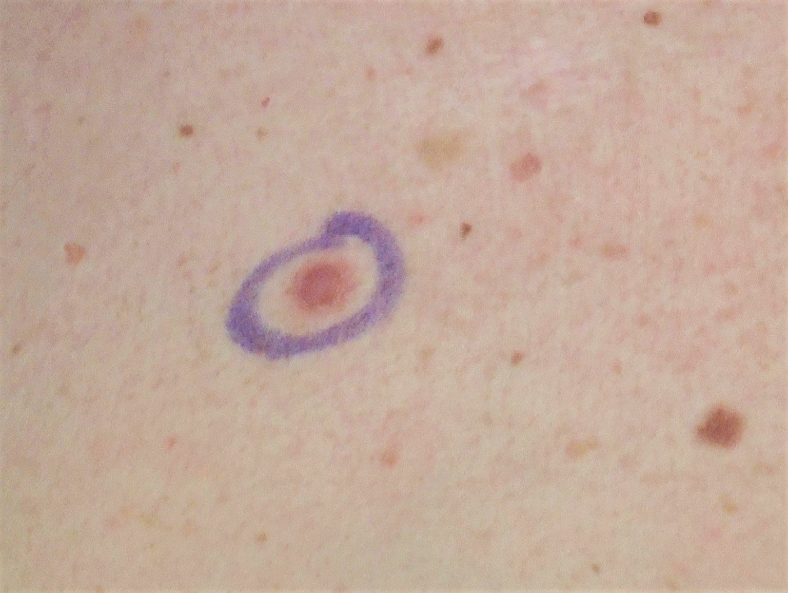
MYTH:  Skin cancer always looks like a dark spot.

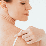
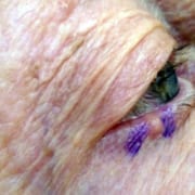
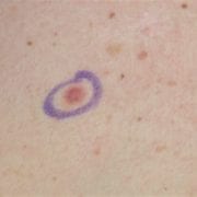
FACT: Skin cancer can have many different appearances. Skin cancer can sometimes look like a pink patch of skin, a pearl-like bump, a scaly patch, a bleeding bump, or a sore that will not heal. Sometimes, skin cancer can also be a dark, raised, or irregular spot. If you notice any of these changes on your skin, please make an appointment to see us.

MYTH: Melanoma is the only type of skin cancer.

FACT: There are multiple types of skin cancers. The most prevalent types of skin cancers are basal cell carcinoma, squamous cell carcinoma, and melanoma. Basal cell carcinoma is the most common type of skin cancer. Melanoma is the most serious type of skin cancer. In the United States, it is estimated that melanoma will cause more than 8,400 deaths in 2025.

MYTH: Skin cancer is not treatable.

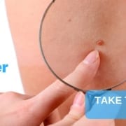
FACT: If caught early, most skin cancers can be highly curable. In fact, patients diagnosed with melanoma who are treated before the melanoma spreads to the lymph nodes have a five-year survival rate of 99%. Regular skin checks at a dermatology practice are important for early detection and treatment of skin cancers.

MYTH: Skin cancer can only occur on sun-exposed areas.

FACT: Skin cancer can occur anywhere on the skin. It can occur under the nails, on the palms of the hands, on the soles of the feet, and on the genitalia. However, the most common areas where skin cancer can develop are on sun-exposed locations, including the scalp, face, ears, neck, chest, arms, and legs.

MYTH: Skin cancer only occurs in older people.

FACT: Among adolescents and young adults aged 15-29 years old, melanoma is the second most commonly diagnosed cancer. Men aged 49 years old and under are more likely to develop melanoma than any other cancer, except for colon and rectal cancers. Women aged 49 years old and under have a higher probability of developing melanoma than any other type of cancer, except breast and thyroid cancers. It is important to have regular skin examinations at a dermatology practice so skin cancers can be detected and treated early.

MYTH: Make-up provides adequate sun protection.

FACT: Some make-up does contain sunscreen, but make-up usually does not provide enough protection from the harmful Ultraviolet (UV) rays of the sun. Sunscreens should be broad-spectrum with coverage against both UV-A and UV-B and at least SPF 30. Sunscreen also needs to be applied multiple times a day for it to be effective. In fact, sunscreen should be applied every two hours, even if the sunscreen is a high SPF.

MYTH: Sunscreens never expire.

FACT: According to the Food and Drug Administration (FDA), sunscreens lose their efficacy and are not as strong after three years. Please always check the bottle for the expiration date and replace sunscreens as needed.

MYTH: It is only necessary to wear sunscreen on sunny, hot days at the beach.

FACT: Sunscreen should be worn every day, even on cloudy and cold winter days.

Clouds can reduce UV rays by only 50%. The remaining UV can still cause sun damage to the skin. And remember to apply sunscreen when at high altitudes, for example, when hiking or skiing. UV increases by 6% for every 1000 feet of elevation gain. When skiing, snow reflects UV rays.

MYTH: Tanning beds are safer than tanning at the beach.

FACT: There is no such thing as safe tanning, whether it is at the beach or a tanning salon. Using a tanning bed before the age of 35 can increase the risk of melanoma by 75%. Also, getting a base tan does not prevent sunburn. All tanning causes DNA damage in the skin, which increases the risk of getting skin cancer.


These myths are just some of the misconceptions about skin cancer. With summer right around the corner and more time spent outdoors, it is important to remember that early detection is key to treating skin cancer. If you notice any changes on your skin that do not resolve within six weeks, please make an appointment with one of the providers at the Dermatologic Surgery Center of Washington. Have a wonderful summer and remember your hats and sunscreen!


Resources

10 Skin Cancer Myths Debunked. University of Texas MD Anderson Cancer Center.  Accessed April 14, 2025. Retrieved from https://www.mdanderson.org/publications/focused-on-health/10-skin-cancer-myths-debunked.h14-1592991.html.

10 Surprising Facts About Indoor Tanning. American Academy of Dermatology. April 26, 2023.  Accessed April 14, 2025. Retrieved from https://www.aad.org/public/diseases/skin-cancer/surprising-facts-about-indoor-tanning.

May is Skin Cancer Awareness Month. American Academy of Dermatology. April 13, 2024. Accessed April 14, 2025. Retrieved from https://www.aad.org/public/public-health/awareness-campaigns/skin-cancer-awareness-month.

Skin Cancer Facts and Statistics. Skin Cancer Foundation. February 2024. Accessed April 16, 2025. Retrieved from https://www.skincancer.org/skin-cancer-information/skin-cancer-facts/.

Skin Cancer. American Academy of Dermatology. February 25, 2025.  Accessed April 14, 2025. Retrieved from https://www.aad.org/media/stats-skin-cancer.

The Most Common Misconceptions About Skin Cancer.  American Society for Mohs Surgery. February 15, 2024. Accessed April 14, 2025. Retrieved from https://www.mohssurgery.org/blog/skin-cancer/the-most-common-misconceptions-about-skin-cancer/

Top 5 Myths About Sunscreens.  Mayo Clinic. May 26, 2023. Accessed April 14, 2025. Retrieved from https://www.mayoclinichealthsystem.org/hometown-health/speaking-of-health/myth-or-fact-coconut-is-an-effective-sunscreen

Weir, H. K., Marrett, L. D., Cokkinides, V., Barnholtz-Sloan, J., Patel, P., Tai, E., Jemal, A., Li, J., Kim, J., & Ekwueme, D. U. (2011). Melanoma in adolescents and young adults (ages 15-39 years): United States, 1999-2006. Journal of the American Academy of Dermatology, 65(5 Suppl 1), S38–S49. https://doi.org/10.1016/j.jaad.2011.04.038


Global Skin Cancer Awareness Month

By Joan Pohutsky, DNP

It’s springtime in the District and the city is coming alive again! Residents are bidding farewell to the cold winter and welcoming the rejuvenating spring season. With this transition comes the month of May, synonymous with blossoming flowers. In the medical community, May also holds a more significant meaning as Skin Cancer Awareness Month.

Spring symbolizes renewal, growth, and the ability to rejuvenate and heal -similar to the natural abilities of human skin. Much like blossoming flowers, human skin cells divide and grow to shield us from external factors. When this growth goes awry, this can lead to conditions like skin cancer.

The Science Behind Your Suntan

As we venture outdoors more often to enjoy the beauty of spring, you might be tempted to work on a suntan. Before you do, however, let’s define what a suntan really is: an injury to the skin in response to two types of ultraviolet radiation (UV). Both types of ultraviolet rays damage the skin and can lead to skin cancer.

  • UV-A rays penetrate deeper into the skin to accelerate signs of skin aging such as wrinkles, sunspots, and leathery skin.
  • UV-B rays are responsible for sunburn. When skin is exposed to UVB rays, the outermost layer (the epidermis) thickens and produces excess melanin, causing the skin to darken in an attempt to prevent further damage to the vulnerable layers below.

Tanning Myths

There are several myths that should be dispelled about tanning:

  1. Tanning is NOT healthy.
  2. Tanning does NOT prevent sunburn.
  3. Tanning does NOT decrease the risk of skin cancer.

What’s the truth about tanning?

  1. Tanning DOES cause skin cancer.
  2. Tanning DOES increase wrinkles, dark spots, and other signs of aging.
  3. Tanning is a sign of injury to the skin and there is NO benefit to a “base tan.”

Supplements and Skin Health

Besides reducing your sun exposure by wearing sunscreen, seeking shade, and covering up, what else can you do to help prevent skin cancer?

Emerging research suggests certain nutrients may play a pivotal role in reducing risks of developing skin malignancies.

  • Nicotinamide (Vitamin B3): supplementation is associated with an overall lower rate of new nonmelanoma skin cancers and actinic keratoses (pre-skin cancers). Notably, a new study recently found that higher doses of similarly structured niacin can increase overall cardiovascular risks including irregular heartbeat and vascular inflammation. As holds true to considering any new medication or supplementation, we recommend that you consult with your primary care physician prior to beginning to use.
  • Antioxidants (Vitamins A, C, & E, plus selenium, polyphenols, lycopene, beta-carotene): prevent and slow damage to cells caused by free radicals, thus neutralizing effects of UV radiation (found in fruits and vegetables). Polypodium Leucotomus is one such antioxidant derived from a fern and plays a role in reducing inflammation and DNA damage in skin cells, thus reducing the risk of skin cancer and photoaging. Polypodium Leucotomus is the active ingredient in Heliocare supplementation.
  • Vitamin D: plays a role in controlling abnormal multiplication of skin cells (found in fortified dairy products, fatty fish, and supplements). While the sun is a natural source of vitamin D, it is safer and more reliable to obtain this from diet and supplementation due to prolonged sun exposure increasing cancer risk.

Nutrients and supplements can influence the overall health, aging, and appearance of our skin.

  • Omega 3 supplementation (fish, nuts, seeds, and vegetables): strengthens skin membranes to prevent moisture loss, has anti-inflammatory properties to reduce redness and swelling, and can aid in the healing process when skin is damaged.
  • Zinc (found in meat, fish, and seafood): acts as a physical barrier to reflect UV rays and decreases inflammation, thus playing a role in wound healing and skin integrity.
  • Metformin: a drug classically utilized for patients with type 2 diabetes, has antiaging properties including decreasing chronic inflammation and oxidative stress. There are observational studies to suggest that long-term use can be associated with reduced skin cancer risk.

A healthy diet and lifestyle contribute to skin health, but sun protection remains the cornerstone of skin cancer prevention. This includes applying broad spectrum sunscreens SPF 30 or higher and reapplying every two hours, seeking shade during peak sunlight hours, and wearing ultraviolet protection factor (UPF) rated sun clothing as well as wide-brimmed hats and sunglasses. Check your skin for new, changing, or suspicious lesions, and enjoy everything spring has to offer!


Sources

Chen A, Martin A, Choy B, et al. A Phase 3 Randomized Trial of Nicotinamide for Skin-Cancer Chemoprevention, 2015; New England Journal of Medicine; N Engl J Med 2015;373:1618-1626; DOI: 10.1056/NEJMoa1506197

Cruz RYMS, Arévalo SV, Rashid A, Jara MRA, Prado MSA. Antioxidant and photoprotective potential of Polypodium leucotomos. Explor Med. 2022;3:607–16. https://doi.org/10.37349/emed.2022.00117

Dong Y, Wei J, Yang F, Qu Y, Huang J, Shi D. Nutrient-Based Approaches for Melanoma: Prevention and Therapeutic Insights. Nutrients. 2023; 15(20):4483. https://doi.org/10.3390/nu15204483

El Mansouri MEssaddouki SMouradi M, et al. Evaluation of the effectiveness and safety of combined oral and topical photoprotection with a standardized extract of Polypodium leucotomos (Fernblock®)Photodermatol Photoimmunol Photomed202339607612. doi:10.1111/phpp.12904

Ferrell, M., Wang, Z., Anderson, J.T. et al. A terminal metabolite of niacin promotes vascular inflammation and contributes to cardiovascular disease risk. Nat Med 30, 424–434 (2024). https://doi.org/10.1038/s41591-023-02793-8

Michalak M, Pierzak M, Kręcisz B, Suliga E. Bioactive Compounds for Skin Health: A Review. Nutrients. 2021; 13(1):203. https://doi.org/10.3390/nu13010203

Sahdeo Prasad, Subash C. Gupta, Amit K. Tyagi, Reactive oxygen species (ROS) and cancer: Role of antioxidative nutraceuticals, Cancer Letters, Volume 387, 2017, Pages 95-105,

ISSN 0304-3835, https://doi.org/10.1016/j.canlet.2016.03.042. https://www.sciencedirect.com/science/article/pii/S0304383516302026

Sami Raza, Firas Al-Niaimi, Faisal R Ali, A systematic review of the uses of metformin in dermatology, Clinical and Experimental Dermatology, Volume 48, Issue 2, February 2023, Pages 73–79, https://doi.org/10.1093/ced/llac027

How to Prevent Skin Cancer.  American Academy of Dermatology (2024). Retrieved from

https://www.aad.org/public/diseases/skin-cancer/prevent/how

How to Properly Examine Skin for Signs of Cancer

Peace of Mind During Pandemic

May is Melanoma Awareness Month, and while you don’t need another thing to worry about these days, it’s important to learn about melanoma and understand its risks. Approximately 200,000 melanoma cases will be diagnosed in the U.S. in 2020 and it’s estimated that the number of new melanoma cases diagnosed will increase by almost 2 percent this year. Men age 49 and under have a higher chance of developing melanoma than any other cancer, and women age 49 and under are more likely to develop melanoma than any other cancer except breast and thyroid cancers.

The good news is that the vast number are curable, especially when diagnosed in the earliest stages. The estimated five-year survival rate for patients whose melanoma is detected early is about 99 percent.

How to Check Your Skin for Cancer

You can stop worrying about melanoma by taking a good look at your skin once a month. Look for anything new, changing or unusual on both sun-exposed and sun-protected areas of the body. Melanomas commonly appear on the legs of women, and the number one place they develop on men is the trunk. Keep in mind, though, that melanomas can arise anywhere on the skin, even in areas where the sun doesn’t shine, including the groin, genitalia, the bottom of your feet and in or around your nails.

What to Look for During Skin Screening

I recommend comparing a suspicious spot to surrounding moles to determine if it looks different from its neighbors. These “ugly ducklings,” or outlier lesions, can be larger, smaller, lighter or darker compared to surrounding moles. A solitary lesion without any surrounding moles for comparison are also considered ugly ducklings. About 20 to 30 percent of melanomas develop in existing moles, while 70 to 80 percent arise on seemingly normal skin.

Not every melanoma is dark or pigmented. Amelanotic melanomas are missing the dark pigment melanin that gives most moles their color. These melanomas may be pinkish, reddish, white, the color of your skin or even clear and colorless, making them difficult to spot.

Look out for any new moles or freckles that arise on your skin, a sore or spot that does not heal, a change in any existing mole (growing, swelling, itching) or any spot, mole or lesion that looks unusual.

You might find lots of things that suggest a lesion of concern, but another encouraging bit of information is that many lesions that meet these criteria are benign, and can be diagnosed while you are in the comfort of your own living room.

Non-invasive Biopsy from Home

Through telemedicine, I see many dark spots that are actually benign. Seborrheic keratoses, benign sun spots, and even ticks can look like melanoma. During this past month I am gratified to be able to reassure patients who are concerned about new lumps and bumps. Only on a few occasions have I determined that a spot needs further evaluation. What happens then? Does that mean you need to come into the office? Not necessarily.

DermTech PLATechnology has advanced to a degree that we can assess a pigmented mole through an innovative genomic test that can be done by you at home. The DermTech Pigmented Lesion Assay (PLA), which comes in a kit sent to a patient’s home, can determine if a mole is benign or if it has the genomic risk factors of melanoma and needs to be biopsied. The PLA has an adhesive patch that sticks to a mole like tape and is peeled off painlessly to collect skin cells. When administering the test, you draw a circle on the tape to highlight the location of the lesion (as seen in the picture). Skin cells that stick to the adhesive patch are analyzed in a specialized laboratory where the cells are examined for two genes that can indicate the presence of melanoma. This gene expression analysis enables DermTech to accurately distinguish between melanoma and non-melanoma.

With the PLA, there is a less than 1% chance of missing melanoma. It results in fewer unnecessary biopsies, and allows us to limit in-office visits. Plus, it’s painless and doesn’t leave a scar.

What’s the process?

Schedule a telehealth appointment by calling our office at (301) 652-8081 to discuss your skin cancer concerns. If it’s advised that you need to have any lesions further evaluated, we may suggest the DermTech PLA. A kit is sent to your home and we supervise the application of the adhesive sticker via a telehealth visit. Results are available in approximately one week, which means that peace of mind, a rare commodity these days, might just be a week away.

What do Eyes Have to do With It?

R-lower-eyelid-BCC-845x321

You might not think of an eye exam as part of your annual skin check visit to the dermatologist, but there are several serious conditions, including cancers of the eyelids, that can be easily missed without a thorough examination. Your annual skin check should include a visualization of the upper and lower eyelids for tumors, assessment of hair distribution along the lash line to check for alopecia, as well as evaluation of eye symptoms, such as eye irritation or a gritty sensation, which can be associated with skin conditions like rosacea.

If a suspicious skin growth is found on or near the eyelid region, a biopsy should be performed to determine the type of lesion present. The most common malignant growths of the eye area are basal cell carcinoma and squamous cell carcinoma. Other less common types of eyelid cancers are sebaceous carcinoma, Merkel cell carcinoma, and intraocular melanoma. Risk factors for eyelid skin cancers include fair skin (blue eyes and red or blonde hair), immunosuppression, and history of previous radiation to the face.

Types of eyelid skin cancers

Basal Cell Carcinoma (BCC): The eyelid skin is extremely vulnerable to damage from the sun’s ultraviolet rays, making non-melanoma skin cancers, like BCC, quite common. While BCCs rarely spread to the bloodstream, they can grow large and cause disfigurement and sometimes even loss of the eye; this necessitates prompt removal with Mohs surgery, which is sometimes performed in consultation with an oculoplastic surgeon. To prevent BCCs of the eyelids, I recommend that you wear UV blocking sunglasses and don a hat — the wider the brim the better. And of course, always wear sunscreen. Stick Sunscreens for sensitive skin are generally well-tolerated around the eyes.

Squamous Cell Carcinoma (SCC): SCCs make up a smaller portion of eyelid cancers, but have a slightly higher risk of metastasis than BCCs. Prevention and treatment of eyelid SCC is similar to BCC.

Melanoma: Intraocular melanoma is the most common type of cancer that can develop within the eye in adults, yet it is still very rare. It can be found in the back of the eye in the pigmented layer of the eyeball, known as the uvea. This area is not visible to eye doctors during a regular eye exam, therefore it is crucial for individuals with a personal or strong family history of melanoma to undergo yearly dilated eye exams with an ophthalmologist. It is unclear how intraocular melanoma develops, although there are individuals who are genetically predisposed. Anyone with light hair and eye color is at a higher risk of developing eye melanoma, as are welders — either through increased occupational ultraviolet radiation exposure or other environmental causes. Melanoma is a serious diagnosis due to its ability to spread to the bloodstream and other organs. Treatment of intraocular melanoma may include surgery, radiation, and chemotherapy.

Bottom line: Check your eyelids for new growths and protect your eyes from UV exposure. Your eyes are not impervious to skin cancer!


References

Bain, J. (September 25, 2018). Focus on Eyelid Skin Cancers: Early Detection and Treatment. The Skin Cancer Foundation. Available online at: https://www.skincancer.org/blog/eyelid-skin-cancers/

Katella, K. (July 24, 2018). For Eye Cancer, Exams are Crucial. Yale Medicine. Available online at: https://www.yalemedicine.org/stories/eye-cancer-prevention/

Mayo Clinic Staff (September 18, 2018). Eye Melanoma. Available online at: https://www.mayoclinic.org/diseases-conditions/eye-melanoma/diagnosis-treatment/drc-20372376.

Early Detection Just Got VERY High Tech

FotoFinder Automated Total Body Mapping Studio

“Early detection” is something I talk about a lot.  That is for good reason: skin cancer is not only the most common form of cancer in the United States, it is also the most curable – that is, IF it is detected early.  This is why I talk just as much about the importance of regular full body exams.

May is Skin Cancer Awareness Month, so this is an especially good time to mention a powerful new early detection tool I’ve just added to my practice:  FotoFinder.  This breakthrough technology is truly one of the most impactful medical devices I have seen in my career.

FotoFinder combines the very best photographic equipment and German robotic engineering in a way that superbly augments the dermatology professional during full body exams.

Here’s how it works: The computer-controlled machine captures highly detailed photographs of the entire skin surface and merges them into a comprehensive Total Body Map.  It then employs artificial intelligence to meticulously compare current skin conditions to earlier “baseline” images.  If it “sees” new moles, or even minute changes in existing moles, it automatically flags them for detailed follow up by the professional.  The mapping and analysis process take just 20 minutes.  It is amazing technology that will greatly advance early detection of skin cancer.

Baseline: FotoFinder Total Body Map

Follow Up: Updated Body Map, with moles highlighted for further examination

When I saw this sophisticated device in action, I knew immediately I need to bring this to our patients.  The Dermatologic Surgery Center of Washington is the first and only practice in the Washington, DC region to install this significant advancement in the science of early detection.

Since we installed the system in January, I am often asked who should consider FotoFinder early detection technology?  If you can answer yes to any of these questions, I recommend we discuss if FotoFinder is right for you:

  • Do you have a history of melanoma or other skin cancer?
  • Do you have a family history of skin cancer?
  • Do you have many moles – more than 50?
  • Do you have large moles – 2 inches in diameter or larger?
  • Do you see changes in a mole or have new moles?
  • Do you have a history of a severe, blistering sunburn as a child or adolescent?
  • Do you have very light skin?
  • Are you exposed to strong sunlight on a regular basis?

Skin Cancer Awareness Month is a great time to make an appointment with one of our talented dermatology professionals for your next full body exam – and create your baseline Total Body Map with FotoFinder.

Click here to make an appointment online or call us at 301-652-8081.


All the photos used are courtesy: FotoFinder Systems, Inc.

Test your skin cancer IQ

Skin cancer is the most common cancer in the United States, and it is estimated that one in five Americans will develop this condition in their lifetime. Skin cancer is triggered by mutations or defects in DNA, leading to cells growing out of control to form malignant tumors.

A Breakthrough In Melanoma Detection

Are you reluctant to undergo a skin biopsy because of the potential for a scar? We have good news. There is a new, non-invasive melanoma detection method to check suspicious dark moles. Read the story below by Dr. Maral Skelsey and learn more about DermTech, a “bladeless biopsy.”

What is Seborrheic Keratoses?

Have you ever been concerned about a dark mole on your skin, only to be told by your dermatologist that it’s not a mole but a seborrheic keratosis? We might tell you not to worry about it, but did you get a good explanation of what they are?

A seborrheic keratosis (SK) is a very common skin growth caused by a thickening of an area of the top skin layer. It usually appears as a brown, black, or light tan growth on the face, trunk or arms, and may have a waxy or scaly raised appearance. Despite the way they look, these growths are harmless. They do not usually cause any symptoms, but they may itch.

Treatment Options for Seborhheic  Keratosis

Treatment of SKs is usually not necessary, but may be desired for cosmetic reasons. Under local anesthesia, they may be removed with a scalpel, scraped off with a special instrument, or burned off with an electric current. The most common treatment for SKs is liquid nitrogen therapy. However, all of these treatments have the potential to leave behind a white mark or  small scar. 

Are seborrheic keratoses contagious?

No. Although they have a warty appearance, these lesions are not caused by a virus and are not contagious.

Are seborrheic keratoses cancerous?

No. SKs may look like pre-cancerous skin growths such as actinic keratoses and have the dark irregular appearance of melanoma, these growths are benign.

Who gets seborrheic keratoses?

Anyone can get these growths- especially anyone with fair skin and those with family members with seborrheic keratoses. People with darker skin types tend to get smaller seborrheic keratoses on the face. Pregnant women may also get them

Do children get seborrheic keratoses?

No. Seborrheic keratoses typically occur in middle-aged and older adults. Think of them as a sign of wisdom!

Contact us with any other questions or to make an appointment with a dermatologist.

Amelanotic Melanoma: Symptoms, Causes, Treatment

melanomaA patient recently came to the office concerned about a new dark mole on her leg. During her full body skin examination, I noticed a pink bump on her back.  I asked her about it and she said it was a bug bite that was taking a long time to heal.  Because of the appearance of the lesion when I examined it with a dermatoscope, I recommended a biopsy. Unfortunately, the pink spot on her back turned out to be a melanoma. The mole on her leg was benign.


Most patients are concerned when they spot a new dark mole, but it’s not widely recognized that melanoma can present as a pink or colorless skin lesion. Amelanotic melanoma is a serious form of skin cancer where the cells do not make melanin or pigment. It is responsible for approximately 2-20% of melanoma cases1. Because of their lack of color, diagnosis of this type of melanoma may be delayed until it reaches an advanced stage.

Symptoms

One of the most obvious symptoms of amelanotic melanoma is the sudden appearance of a red, pink, or skin-colored spot on your body where it wasn’t before. Melanomas also grow and may change shape, becoming a larger bump over time. Amelanotic melanoma may not present with the classic ABCD criteria (Asymmetry, Border Irregularity, Color variation, and Large Diameter) that are typically associated with melanoma warning signs. Expanding the ABCD warning signs to include the 3 Rs (Red, Raised, Recent change) may help in the early detection of amelanotic melanoma2. It is also estimated that about 25% of melanomas of the nail are amelanotic, so checking your nails should be a regular part of self-skin examinations. It is important to note that melanoma may present differently in individuals of color, therefore regular self-skin examinations are essential in the early detection of these skin cancers.

Causes

Melanoma occurs when the DNA in your skin cells becomes damaged, which can cause the cells to grow out of control and become cancerous. Exposure to the sun for long periods of time can damage your skin cells and increase your risk for melanoma. This risk is increased in patients with fairer skin, individuals of European descent, those with many moles (especially 50 or more), a family history of melanoma, and patients with a weakened immune system from an existing condition3. The use of tanning beds has also been associated with an increased risk for melanoma and should be avoided4.

Treatment

The most common treatment for melanoma is surgical removal.  However, melanoma can spread to the lymph nodes, necessitating the removal of one or more nodes. Advanced melanoma may need to be treated with chemotherapy and/or radiation. Biological therapy (or drugs that aid your immune system in killing cancer cells) as well as targeted therapy (medications that weaken cancer cells) are other common melanoma therapies. Patients with a history of a previous melanoma are 4 times more likely than the general population to develop a subsequent invasive melanoma, therefore full body skin examinations should be performed by a dermatologist every 3 months after a melanoma diagnosis.


References:
1.         Thomas, N.E., Kricker, A., & Waxweiler, W. Comparison of Clinicopathologic Features and Survival of Histopathologically Amelanotic and Pigmented Melanomas: A Population-Based Study. JAMA Dermatol. 2014;150(12):1306-1314. doi:10.1001/jamadermatol.2014.1348
2.         Kim, S.J., Park, H.J., Lee, J.Y., Cho, B.K., A Case of Subungual Melanoma. Ann Dermatol. 2008 Mar; 20(1): 26–28. Published online 2008 Mar 31. doi:  10.5021/ad.2008.20.1.26
3.         American Cancer Society. Risk Factors for Melanoma Skin Cancer. Available online at: https://www.cancer.org/cancer/melanoma-skin-cancer/causes-risks-prevention/risk-factors.html
4.         American Academy of Dermatology. Indoor Tanning. Available online at: https://www.aad.org/media/stats/prevention-and-care